
Muscle Fat Accumulation Emerges as a New Aging Indicator
Why Muscle Fat Matters in Aging Research
Scientists have identified increasing fat deposits within skeletal muscle as a promising marker for assessing biological age. The discovery, announced at a recent conference, could transform how researchers evaluate age‑related decline across populations.
Health news
 Vegan Diets May Reduce Gut Bacteria Diversity, Study Finds
 Music with beats cuts stress hormone more than pink noise
 Epigenetic Shifts Can Trigger Liver Cancer Without DNA Damage
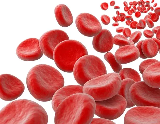 Blood Clots and Brain Tumors Linked by Overactive Platelets
Traditional aging metrics rely on chronological age or broad health surveys. Researchers now focus on cellular and tissue changes that directly reflect physiological wear. Fat infiltration, or myosteatosis, occurs when lipid droplets accumulate in muscle fibers, reducing strength and metabolic function. By measuring this process, scientists hope to quantify the body’s cumulative damage more accurately.
Muscle tissue is central to mobility, metabolism, and overall health. As people age, muscle mass naturally decreases—a condition called sarcopenia. However, the quality of remaining muscle also changes. Fat infiltration disrupts muscle fiber integrity, leading to weaker joints and slower recovery times. Dr.
Elena Marquez, a gerontologist at the Institute for Aging Studies, explained, „When we see fat within muscle, it signals that the body’s repair mechanisms are faltering. It’s a tangible sign of biological aging.”
Can Imaging Turn Fat Infiltration into a Practical Tool?
Studies across diverse age groups have shown a strong correlation between the extent of intramuscular fat and functional decline. In one longitudinal cohort, participants with higher fat levels experienced a 15% greater risk of falls over five years. These findings suggest that measuring fat infiltration could predict future health events more reliably than age alone.
Magnetic resonance imaging (MRI) and ultrasound are the primary techniques used to quantify muscle fat. MRI offers precise volumetric data but is costly and less accessible. Ultrasound, meanwhile, provides rapid, bedside assessments, though it requires operator skill.
Researchers are working on standardizing protocols so that clinicians can routinely screen patients for myosteatosis.
What Does This Mean for Public Health?
„We’re developing algorithms that translate ultrasound images into simple fat‑in‑muscle scores,” said Dr. Marquez. „Once validated, these scores could be added to routine check‑ups, giving doctors a clear picture of a patient’s biological age.”
Identifying fat infiltration early could guide interventions such as resistance training, nutritional adjustments, and pharmacological treatments aimed at preserving muscle quality. Public health programs might incorporate muscle fat screening to target at‑risk groups before significant disability occurs.
Moreover, as societies age, healthcare systems face increasing costs related to frailty and chronic disease. A reliable biomarker like muscle fat infiltration could help allocate resources more efficiently, focusing preventive care on those most likely to benefit.
Frequently Asked Questions
Q1: How is muscle fat measured? Answer: Muscles are scanned with MRI or ultrasound to detect lipid deposits.
The resulting images are analyzed to calculate the proportion of fat within the muscle tissue.
Q2: Can lifestyle changes reduce muscle fat? Answer: Yes. Regular resistance exercise and adequate protein intake can lower intramuscular fat and improve muscle strength.
Q3: Is this biomarker ready for clinical use? Answer: Standardized protocols are still under development, but pilot studies show promising accuracy for predicting age‑related decline.