
Cone Opsin Structures Reveal New Paths to Treat Vision Disorders
How the Structural Blueprint Advances Vision Science
A team of scientists at the Paul Scherrer Institute in Switzerland, led by structural biologist Polina Isaikina, announced on June 25 2026 that they have mapped the three‑dimensional structures of human cone opsins. The work, published in a peer‑reviewed journal, offers a detailed view of the light‑sensing proteins that power daytime vision and explains how genetic defects can trigger blindness.
Health news
 Sleep May Be the Heart's Nightly Repair Window, Study Suggests
 APOE Genotype Influences How Well Blood Tau Protein Predicts Cognitive Decline
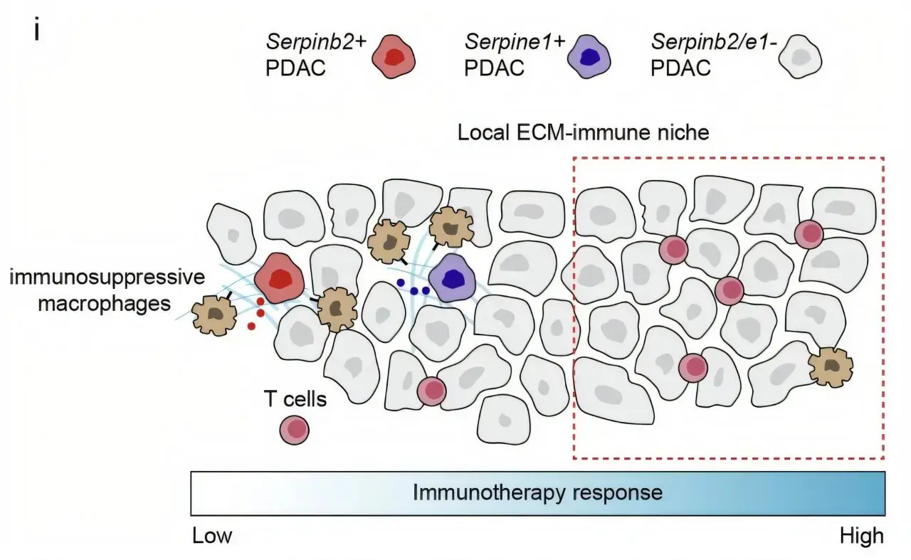 Pancreatic Cancer Hijacks Blood-Clotting System to Evade Immune Attack
 Survey Reveals Most Men Unaware Dairy May Increase Prostate Cancer Risk
The researchers used cryogenic electron microscopy to capture cone opsins in both inactive and light‑activated states. By comparing the shapes, they identified the molecular switches that open and close the protein’s retinal binding pocket. Mutations linked to achromatopsia and other cone‑degeneration disorders were found to disrupt these switches, causing the proteins to misfold or lose stability. „Seeing the protein at atomic resolution lets us pinpoint exactly where disease‑causing changes occur,” Isaikina said.
The new models show that cone opsins share a common scaffold with rod opsins but possess unique loops that fine‑tune color perception. The team reconstructed the proteins from the three cone types—S, M and L—that detect short, medium and long wavelengths. Detailed maps of the binding sites revealed how specific amino‑acid substitutions alter spectral sensitivity. Experiments in cultured retinal cells confirmed that restoring the normal shape of the binding pocket rescued light response in cells bearing disease‑associated mutations. This functional validation suggests that small molecules or gene‑editing tools could correct the faulty proteins in patients.
Could These Findings Lead to Therapies for Blindness?
The structural insights open several therapeutic avenues. One possibility is designing pharmacological chaperones that stabilize mutated opsins, preventing them from aggregating. Another is improving gene‑therapy vectors to deliver corrected opsin genes more efficiently. „We now have a molecular blueprint to guide drug design and to test how engineered opsins behave in the eye,” noted co‑author Dr. Marco Keller. Clinical trials for cone‑specific gene therapy are already underway, and the new data may accelerate their progress.
The study marks a milestone in visual‑science research, bridging the gap between genetics and protein chemistry. As scientists translate these structural maps into treatments, patients with inherited cone disorders could soon benefit from therapies that restore color vision and sharpen daylight sight.
Frequently Asked Questions
What are cone opsins and why are they important? Cone opsins are light‑sensitive proteins in cone cells that enable color vision and high‑resolution perception in bright light. Their proper function is essential for daily activities like reading and driving.
How does the new structural information help treat vision loss? By revealing the exact shape and activation mechanism of cone opsins, researchers can design drugs or gene therapies that correct the defects caused by disease‑linked mutations.
When might patients see the results of this research? Therapeutic development typically takes several years. If preclinical studies succeed, clinical trials could begin within the next five to ten years, offering hope for future patients.
Content written by Dr. Elena Voss for wellness-bio-radar.com editorial team, AI-assisted.